

Born to Explore.
Born to Explore.
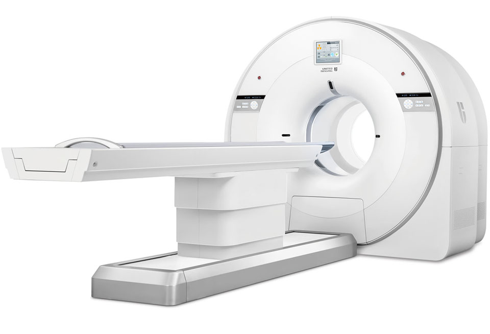
The uBioEXPLORER is a cutting-edge translational digital total-body PET/CT system that boasts an unparalleled 50 cm bore diameter and 48 cm axial PET field of view (FOV), making it possible to capture the entire anatomy of different animals – from rodents to non-human primates – in a single bed position. Utilizing the same technology as our uEXPLORER® clinical total-body PET system, the uBioEXPLORER has the capability to perform total-body dynamic scanning, resulting in ultra-high image resolution and ultra-low radiation doses, thereby revolutionizing the conventional approach to preclinical whole-body PET/CT imaging. The system's extensive coverage and industry-leading sensitivity facilitate the observation of dynamic changes in radiotracer distribution with remarkable temporal resolution, thus enabling translational applications and scientific research to reach new heights with the uBioEXPLORER.
The large 50 cm bore diameter and 48 cm axial PET FOV provides exceptional image quality with 2.6 mm NEMA resolution for preclinical imaging – from rodents to non-human primates.
One Size Fits All
With 94,080 crystal elements, the uBioEXPLORER provides an industry-leading 57 cps/kBq system sensitivity for preclinical total-body PET/CT imaging applications which helps improve quantitative accuracy and small lesion detectability.
Total-Body PET/CT, Miniaturized
Speed is king. The 80-slice, ultra-low noise clinical diagnostic CT built into the uBioEXPLORER provides outstanding CT electronic noise performance to optimize scan speed, dose efficiency, and image quality.
Redefine Clinical and Research Efficiency

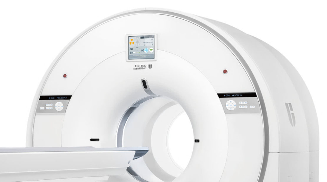
Integrated-Light-Guide Digital PET Detector
The Integrated-Light-Guide improves light collection efficiency and spatial resolution to achieve exceptional image quality.
80-slice CT with Z-Detector
The ultra-low noise design of the Z-Detector helps produce high image quality with low radiation dose.
Ultra-High Sensitivity
With an ultra-high system sensitivity of 57 cps/kBq, the uBioEXPLORER improves the tradeoffs between scan time, radiation dose, and image quality.
Long Axial Field of View
The long 48 cm axial FOV and high system sensitivity of the uBioEXPLORER enables total-body dynamic imaging with fine temporal resolution.
Large Bore Diameter
The large 50 cm bore diameter of the uBioEXPLORER maximizes the versatility of the system for various preclinical imaging applications – from multi-rodent imaging to larger non-human primates.
The uBioEXPLORER preclinical digital PET/CT maintains a stable temperature environment for consistent detector performance with minimal infrastructure investment.
This fully configured 80-slice diagnostic CT produces excellent image quality with a high image resolution for a variety of different protocols.
Unlock the possibilities for translational research utilizing the uBioEXPLORER's exquisite PET and CT resolution, with the highest preclinical PET sensitivity on the market.
Enable new drug discovery by visualizing dynamic total-body PET tracer distribution including blood, organs, and tissues over time in one single bed position.
Swine model whole-body imaging
PET/CT images showing normal 18F-FDG whole body uptake in a 35 kg pig.
Scan protocol: uBioEXPLORER, 3.24 mCi of 18F-FDG, 60 min post-injection, 30 min scan time, 3 scan beds with scan duration of 10 min/bed.
Dynamic FDG distribution visualization in a rabbit model
PET/CT images showing normal dynamic 18F-FDG distribution in a 2.5 kg rabbit.
Scan protocol: uBioEXPLORER, 1.3 mCi of 18F-FDG, data acquired for 60 min and time-binned in a 1 and 3 s frames, data reconstructed using ordered subset expectation maximization (OSEM) with point spread function (PSF) modeling.
Multiple post-injection delay timepoint imaging in a rabbit model
PET images showing multiple post injection delay timepoint whole-body imaging in a 2.5 kg rabbit, allowing accurate assessment of 18F-FDG distribution over time.
Scan protocol: uBioEXPLORER, 1.3 mCi of 18F-FDG, 1 scan bed, 5 min scan duration; 55 min, 7 h and 10.5 h post-injection.
Translational PET/CT imaging in different animal models
PET images showing normal 18F-FDG uptake in a 2 kg rabbit, 12 kg beagle and a 35 kg pig, exhibiting the translational capabilities of the uBioEXPLORER.
Images Courtesy of Shanghai Institute of Materia Medica, Chinese Academy of Sciences.